Cardiac Surgery
There are three major categories of heart surgery. They are the following:
Valve replacement procedures
Coronary artery bypass graft (CABG) procedures
Heart transplant
Surgeons worldwide are rapidly gaining experience with left ventricular reduction (heart reduction surgery) as a treatment for congestive heart failure and an alternative to heart transplantation.
Valve Replacement Procedures
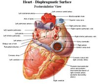
Normal, healthy heart valves open easily, close securely, and do not allow blood flow to return through them once they are shut. There are four heart valves. Two control the inflow of blood to the ventricles from the atria; while the second set controls the outflow of blood from the heart to the body. Each of the heart valves are composed of two or three flaps of fibrous tissue called leaflets. The leaflets act as one-way doors opening to allow the flow of blood in one direction and closing to prevent the blood from backing up into chambers of the heart that it has just left.
The valves between the atria and ventricles (tricuspid on the right side and mitral on the left) have fibrous cords (chordae tendineae) that help the valve's overlapping leaflets to function by connecting them to the muscle wall of the ventricles. The valves that control the flow of blood from the ventricles to the arteries (pulmonary on the right side and aortic on the left) are cup-shaped structures that do not overlap. Other muscles or structures inside the heart do not help these valves.
Common problems with the heart valves include insufficiency due to a leaky valve and torn fibers (chordae tendineae) connecting the valve leaflets to the papillary muscles on the wall of the ventricles. Valve problems that may be present at birth are called congenital malformations. Injury, infection, or illnesses such as rheumatic or scarlet fever may cause problems with the heart valves. Another condition, called valvular stenosis, occurs when the heart valves become thick and stiff, thereby limiting their ability to function properly. The most common heart valve problem is stenosis of the aortic and mitral valves.
During surgery to correct or replace a defective heart valve, the patient is deeply anesthetized and the chest opened. A heart-lung bypass machine under the direction of a perfusionist and the anesthesiologist assumes the flow of blood throughout the body. The pumping action of the heart is stopped to allow the surgeon to make an incision in the heart, granting access to the defective valve. The surgeon will repair the valve or completely replace it with an artificial device. When a replacement device is installed, it is carefully sutured into place and checked before the heart is closed. Following the surgery, the patient will spend time in recovery and possibly an area of the hospital where they can be monitored. In most cases, the patient is ready to return home in a few days.
The success rate for heart valve surgery is high and continues to increase thanks to improved technology and surgical techniques. The operation provides symptom relief, usually improving both quality and quantity of life for the patient. Life-long anticoagulant therapy is required for patients with artificial heart valves. It is not unusual for the clicking of the mechanical heart valve to be heard in the chest.
Coronary Artery Bypass Graft (CABG) Procedures
Coronary artery bypass graft surgery or CABG (pronounced "cabbage") is the most common open-heart procedure. This surgery provides relief to patients who have blocked or narrowed arteries due to atherosclerosis. The symptoms of atherosclerosis or "clogging of the arteries" often include the following:
* Shortness of breath upon exertion
* Chest pains or tightness in the chest
* Dizziness
* Feelings of nausea and sweats
As the coronary arteries continue to narrow, the blood supply to the heart muscle is reduced or blocked altogether. When this happens, the patient experiences a heart attack or myocardial infarction.
Bypass surgery provides alternative routes of blood flow around the narrowed regions of the coronary arteries, detouring the blockage. A blood vessel from another part of the body is sewn into place to route blood around the blockage, restoring normal blood flow to the heart muscle.
The most common source of this new pathway is a vein from the lower extremity called the Greater Saphenous Vein (or GSV), that runs from just inside the ankle bone to the groin. This vein is useful because it is long and straight, and since it is just one of a large series of veins in the legs, it's function may be easily assumed by the other vessels present in the legs. Another major vessel used for bypass grafts is the Left Internal Mammary Artery (LIMA). This vessel lies on the undersurface of the sternum (breastbone), making it easily accessed during surgery. The lower end may simply be detached and connected to one of the coronary arteries on the surface of the heart.
During surgery, the patient is deeply anesthetized and the chest opened. A heart-lung bypass machine under the direction of a perfusionist and the anesthesiologist assumes the flow of blood throughout the body. The pumping action of the heart is stopped to allow the surgeon to make the delicate connections of the bypass graft vessels to the coronary arteries. Depending upon the number and complexity of the grafts, the procedure may take from three to six hours. Following the surgery, the patient will spend time in recovery and then an area of the hospital where they can be monitored.
In most cases, the patient is ready to return home in less than a week. Typically, it takes another two to three weeks for most patients to feel stronger and regain normal body habits, such as appetite, sleep patterns, and bowel action. For patients in non-physical jobs, most can return to work within four to six weeks, depending upon their energy level. After full recovery, the most patients can return to a full and active lifestyle that includes moderate exercise, travel, and employment.
Over 200,000 coronary artery bypass graft procedures are performed annually in the United States alone. The vast majority of these patients have an excellent chance for a full recovery.
Heart Transplantation
With over 1,500 cases per year performed, heart transplants are the third most common transplant operation in the U.S. following cornea and kidney transplant procedures. A health heart is obtained from a donor who has suffered brain death, yet remains on life-support. A surgical team retrieves the healthy heart from the donor and transports it in a special solution that preserves the organ.
The recipient is placed into a deep sleep through general anesthesia. The chest is opened and the patient's blood flow rerouted through a heart-lung machine. The patient's diseased heart is removed and the donor heart stitched into place.
A heart transplant may be recommended for patients experiencing heart failure due to coronary artery disease; cardiomyopathy (thickening of the heart walls); heart valve disease with congestive heart failure; and severe congenital heart disease. Heart transplantation surgery is not recommended for patients who have kidney, lung, or liver disease; insulin-dependent diabetes mellitus; or other life-threatening diseases.
The heart transplant procedure is effective in prolonging the life of a person who otherwise would die due to heart disease. Worldwide, approximately 80% of heart transplant patients are alive two years after the procedure.
Graft rejection is the main post-operative problem facing transplant patients. Immunosuppressive medications must be taken indefinitely. The immune system of the body views the transplanted heart as a foreign body and fights it as though it were a major infection. The immunosuppressive drugs inhibit the body's attempts to reject the donor heart; however, they also weaken the body's ability to defend itself against various common infections.(source:cardioconsult.com)


0 Comments:
Post a Comment
<< Home